

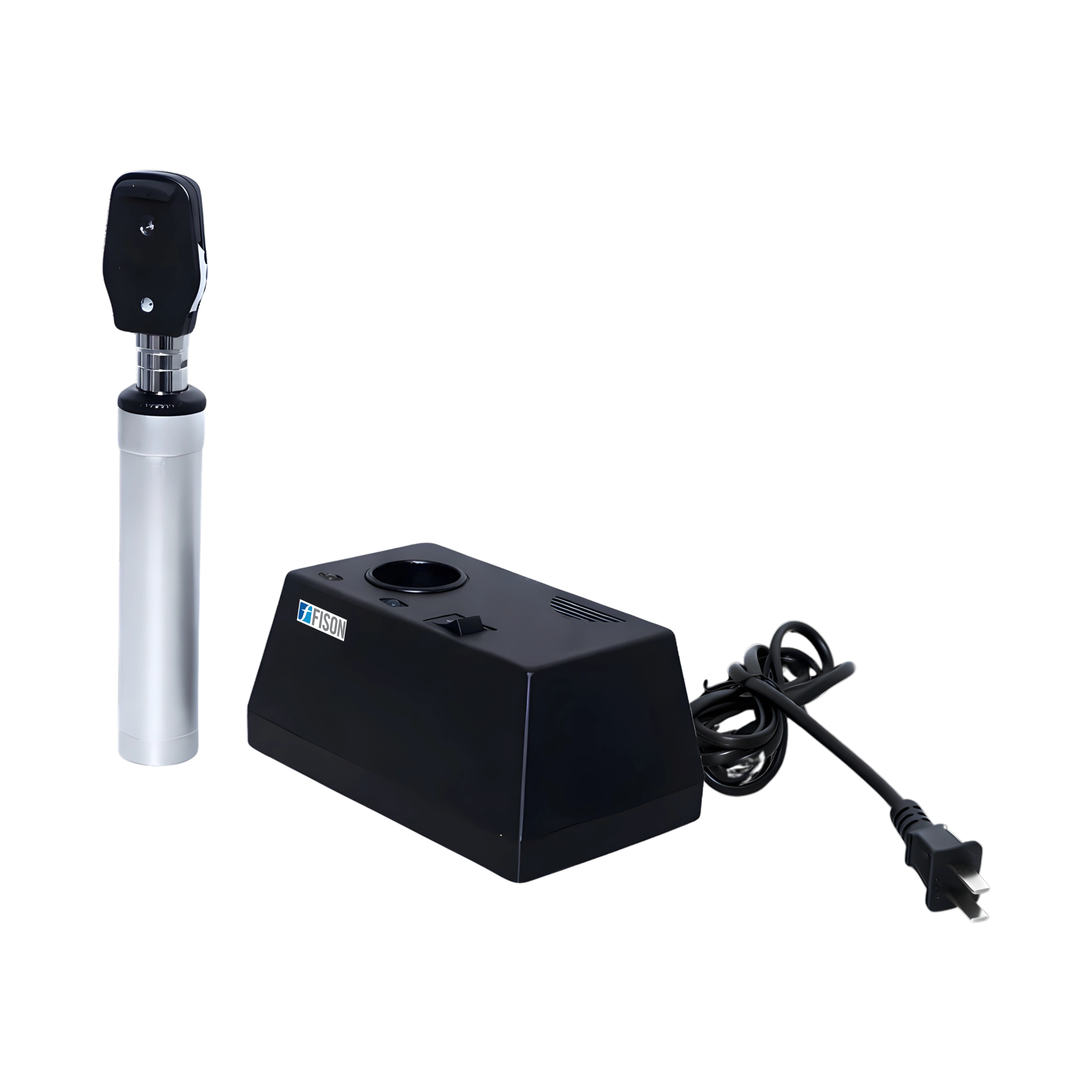
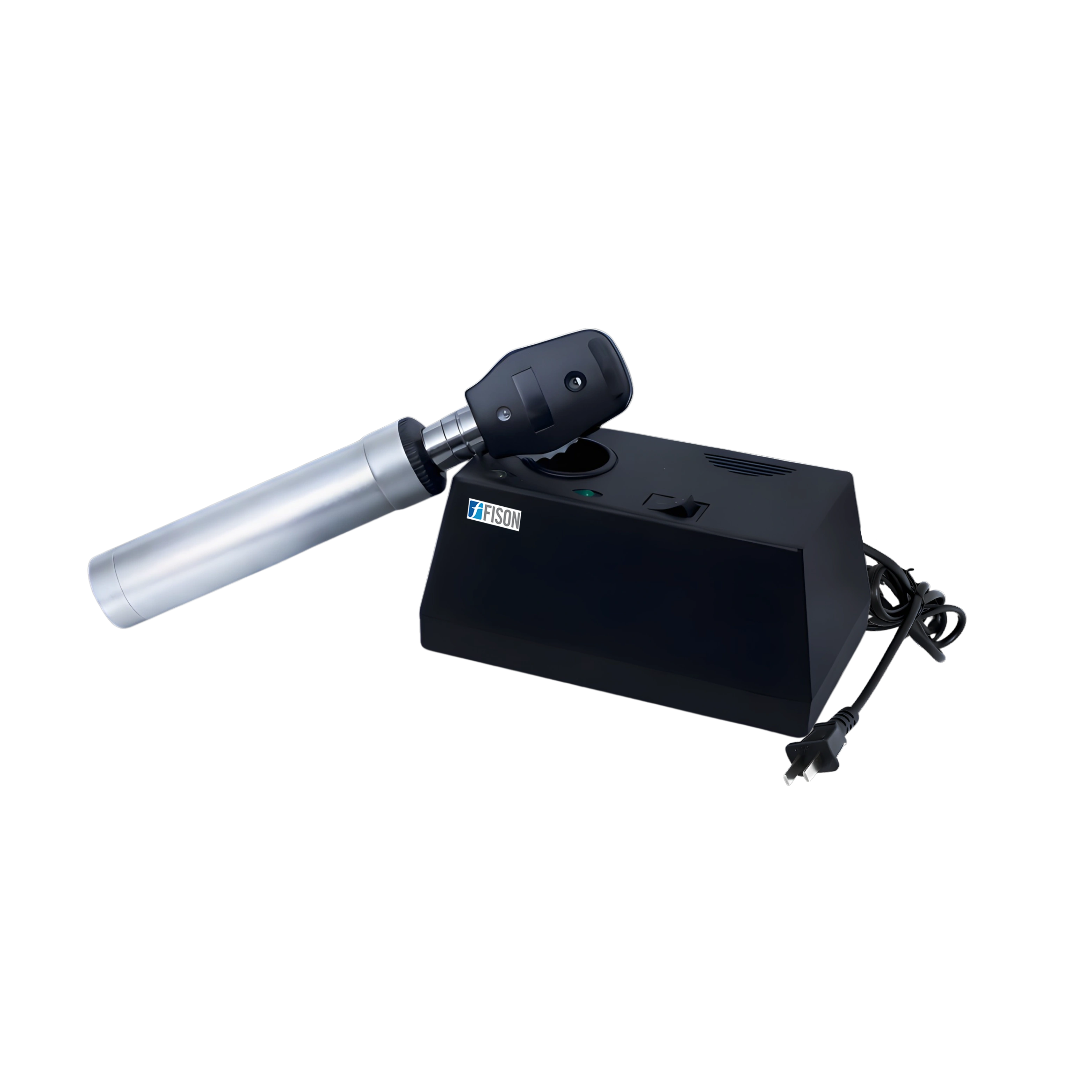
Fison Ophthalmoscope FM-OTS-A100 combines a 3.7 V, 2.6 W mini halogen lamp with illumination options of a large Φ3.2 mm spot, a small Φ1.6 mm spot, a slit and a central net. A red-free filter supports clearer visualization, and diopter compensation from -25D to +20D allows focus adjustment during viewing. This ophthalmic examination equipment allows visual inspection of the interior structures of the eye.
$530.00
| Illumination | large light spot (Φ3.2mm) small light spot (Φ 1.6mm) slit central-net Redfree filter |
| Diopter Compensation | 0, ±1D, ±2D, ±3D, ±4D, ±5D, ±6D, ±8D, ±10D, ±12D, ±16D, ±20D, -25D |
| Light Source | 3.7V, 2.6W Mini-halogen lamp |
| Power Supply | AC 220V ± 10% / 50Hz ± 10%; AC 110V ± 10% / 60Hz ± 10% |
| Input Power | 5VA |
| Weight | 2 Kg (Battery included) |
Ophthalmoscope FM-OTS-A100 is used in ophthalmology clinics, hospitals, and eye care centers to examine the retina and other internal eye structures for detecting and monitoring conditions such as retinal disorders, glaucoma, and cataracts.
Fison Ophthalmoscope provides large and small light spots from ?1.6 mm to ?3.2 mm, along with slit, central-net and red-free filter illumination. Diopter compensation from -25D to +20D lets the examiner bring the retina into focus, and a mini-halogen lamp supplies steady illumination. As ophthalmic examination equipment, it allows direct viewing of the fundus, optic disc and retinal vessels at the selected illumination and diopter setting.